How Does Breast Cancer Look Like In Ultrasound / Breast Cancer Stages Explained By Moose And Doc - A rash isn't the only visual symptom of inflammatory breast cancer.
Dapatkan link
Facebook
X
Pinterest
Email
Aplikasi Lainnya
How Does Breast Cancer Look Like In Ultrasound / Breast Cancer Stages Explained By Moose And Doc - A rash isn't the only visual symptom of inflammatory breast cancer.. It is usually preceded by an injury to the breast from a car accident or sports injury. Ultrasound is not used on its own as a screening test for breast cancer. Other ultrasound findings that suggest breast cancer include: What does breast cancer look like on ultrasound. What does a solid mass look like in an ultrasound breast image?
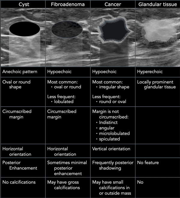
Can be idiopathic, related to steroid abuse or associated with hormonal treatments such as prostate cancer therapies. Physical examination and mammogram can be more accurate in some settings. With ultrasound, the radiologist will probably be trying to get a sense of the internal texture of the breast lesion and surrounding area. By far the most common abnormalities in the breast, which usually present as a lump in the breast are cysts, fibroadenomas, breast cancer and palpable glandular tissue. May present as a retroareolar lump with or without pain.
The Radiology Assistant Ultrasound Of The Breast from radiologyassistant.nl A specialist looks at the ultrasound pictures. Both the mammogram and ultrasound looked fuzzy and gray on the screen and i have no idea. The dye collection in the breast can also look clumpy or appear in a section of the breast, depending on the involvement of dcis. Can ultrasounds miss breast cancer? What does breast cancer look like on a mammogram? This breast cancer ultrasound image shows changes related to breast cancer that are not seen as microcalcifications or a mass or lump. Dcis on mri may create an area of irregular enhancement of the mri dye into the breast. What does breast cancer look like on an ultrasound?:
A breast ultrasound is most often done to find out if a problem found by a mammogram or physical exam of the breast may be a cyst filled with fluid or a solid tumor.
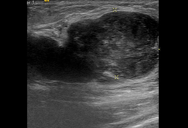
17 years experience general surgery. Solid lesions can be a little brighter or darker than the surrounding tissue, and the way to evaluate these on ultrasound is to look closely at the margins or the outer edges of the nodule. A breast ultrasound is a painless procedure that uses sound waves to make images of the inside of your breast. What does breast cancer look like on a mammogram? Dcis on mri may create an area of irregular enhancement of the mri dye into the breast. Breast cancer is among the most common causes of cancer deaths today, coming fifth after lung, stomach, liver and colon cancers. It is particularly valuable for distinguishing solid from fluid masses, as fluid appears as the darkest material on a sonogram, and solid lesions may appear a little brighter or a little darker than their surroundings. The images are reconstructed as multiple thin slices which can be individually scrolled through to reduce tissue overlap (figs. With ultrasound, the radiologist will probably be trying to get a sense of the internal texture of the breast lesion and surrounding area. Cysts, tumors, and growths will appear as dark areas on the scan. Physical examination and mammogram can be more accurate in some settings. Tumor size is an important factor in breast cancer staging, and it can affect a person's treatment options and outlook. They cause a large amount of the sound waves projected at them to bounce back towards the machine.
It also can be used to look at a suspicious area that was seen on a mammogram. May present as a retroareolar lump with or without pain. You may notice dimpling or pitting, and the skin on your breast. This appears most commonly as streaking, known as linear enhancement. Dcis on mri may create an area of irregular enhancement of the mri dye into the breast.
1 from On ultrasound it will be hypoechoic with spiculations radiating away from the nipple. The images from a breast ultrasound are in black and white. Like standard mammography, tomosynthesis utilizes a paddle to compress the breast to minimize any possible motion as well as minimize the amount of radiation needed to penetrate the breast tissue. Kathleen ruddy inflammatory breast cancer accounts for approximately 5% of all cases of invasive breast cancer in the united states. You can get dressed straight after the ultrasound. By far the most common abnormalities in the breast, which usually present as a lump in the breast are cysts, fibroadenomas, breast cancer and palpable glandular tissue. This appears most commonly as streaking, known as linear enhancement. Cysts, tumors, and growths will appear as dark areas on the scan.
What does breast cancer look like on a mammogram?
It is the most common cause of cancer death in women. in 2005 alone, 519 000 deaths were recorded due to breast cancer. this means that one in every 100 deaths worldwide and almost one in every 15 cancer deaths were due to breast cancer. Cysts, tumors, and growths will appear as dark areas on the scan. A specialist looks at the ultrasound pictures. Dcis on mri may create an area of irregular enhancement of the mri dye into the breast. On ultrasound, a breast cancer tumor is often seen as hypoechoic, has irregular borders, and may appear spiculated. Ultrasound is only one means of evaluation of the breast. A rash isn't the only visual symptom of inflammatory breast cancer. Breast ultrasound is not usually done to screen for breast cancer. The images from a breast ultrasound are in black and white. With ultrasound, the radiologist will probably be trying to get a sense of the internal texture of the breast lesion and surrounding area. This appears most commonly as streaking, known as linear enhancement. Rapid onset of symptoms (redness, swelling, warmth, itching, skin thickening) are hallmarks of the disease. Any area that does not look like normal tissue is a possible cause for concern.
Is the abnormal enlargement of rudimentary male breast tissue. With ultrasound, the radiologist will probably be trying to get a sense of the internal texture of the breast lesion and surrounding area. There is a slight increase in the density in the right breast compared with the left. If a solid lump shows on the scan you might need to have. On ultrasound it will be hypoechoic with spiculations radiating away from the nipple.
Breast Lumps In Young Women Diagnostic Approaches from img.medscapestatic.com My doctor found a lump in my breast during my yearly mammogram and then brought in an ultrasound technician to follow up with an ultrasound for a closer look. Below are images of dcis on breast ultrasound. Mammogram imaging may show skin thickening, but often there is no distinct mass found on physical examination or … Tumor size is an important factor in breast cancer staging, and it can affect a person's treatment options and outlook. What does breast cancer look like on a mammogram? Dcis on mri may create an area of irregular enhancement of the mri dye into the breast. This high amount of echo results in a bright white spot appearing on the ultrasound image. May present as a retroareolar lump with or without pain.
May present as a retroareolar lump with or without pain.
Breast ultrasound is not usually done to screen for breast cancer. We will discuss each of these findings in more detail in a moment. Ultrasound is not used on its own as a screening test for breast cancer. This high amount of echo results in a bright white spot appearing on the ultrasound image. What does breast cancer look like on a mammogram? Dcis on mri may create an area of irregular enhancement of the mri dye into the breast. What does breast cancer look like on an ultrasound?: They cause a large amount of the sound waves projected at them to bounce back towards the machine. Rapid onset of symptoms (redness, swelling, warmth, itching, skin thickening) are hallmarks of the disease. Tumors are likely to be smaller when doctors detect them early, which can. Other ultrasound findings that suggest breast cancer include: Breast cancer is among the most common causes of cancer deaths today, coming fifth after lung, stomach, liver and colon cancers. Learn more about the breast ultrasound procedure, risks, purpose, results, and.
Ideas For Making Teachers Day Card - Greeting Card Ideas Instructions On How To Make Lots Of Handmade Cards / Create the front of the card as previously designed, or just let your imagination flow and make it up as you go along. . Encouragement is fuel for another day of giving and guiding, and every teacher needs that. Diy teacher's day greetings card | handmade teacher's day card making ideas | teacher's day greetings card Teachers day card | teachers day card making | teachers day card easy | teachers day card ideas |hi,welcome to our youtube channel learn with nikitaour play. Decorate the flowers in the ready vase. All we did was print the 'thanks from … Published on may 5, 2016 , under images. To use as seen, or to adapt to their given circumstances with the materials chosen or available. 5th september happy teachers day greeting card. See more ideas about teachers day card, handmade teachers day cards, teacher cards. When your child is making...
What Is The Color For Colon Cancer Awareness - Transparent Colon Cancer Ribbon, HD Png Download ... - The detection and removal of precancerous polyps at awareness is the first step to action. . A guaiac fecal occult blood test (fobt). Colon cancer is the second leading cause of death among cancers. Colorectal cancer screening can catch abnormal cell changes before they become cancerous. What do i do before exam? During a colonoscopy abnormal growths, called polyps, can be removed before they transform into cancers. Colorectal cancer is the second most common cancer diagnosis among females and the third most. Do you know the significance of friday, march 7? Colon cancer often does not cause symptoms in the early stages. Only a medical professional can determine. Colon cancer is one of the most common forms of cancer. Colon Cancer: Cancer Color Meanings from 4.bp.blogspot.com ...
What Sign Are Cancers Most Compatible With : Check Your Star Sign Compatibility Astromatcha / There seemed to be some kind of natural connect already with them. . Taurus, scorpio, virgo and least compatible with aquarius, libra, gemini. Cancers need a home filled with a loving family, and this gives cancer unique compatibility with certain signs of the zodiac. Cancers are loyal and emotional while gemini can be unpredictable. What is the most compatible sign for scorpio woman: There are other planets which also affect someone's personality. Some signs are more compatible with you than others. Everyone has a star sign which actually matches their personality: Unfortunately, every complaint or symptom of cancer can be some cancers occur more frequently in certain age groups. Cancer with cancer is well… a little bit risky. If your rising (personality/ego) and mars (drive/sexuality) signs are in sag, i could very much see most folks not pegging you as a cancer sun. ...


Komentar
Posting Komentar